
Technology & Digital Dentistry at Canobie Dental
Digital scans and 3D images help us explain your options, while in-office crown fabrication can mean fewer visits.
Digital impressions
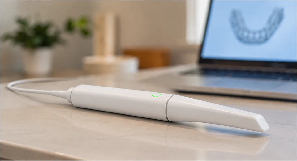
A small intraoral scanner creates a detailed 3D model of your teeth. For many procedures, it replaces a tray of impression material.
The scan gives us a record for crowns, clear aligners, retainers and implant restorations. We can also show you the model as we explain your teeth and treatment options.
Crowns with fewer visits
Digital scans and in-office fabrication let us design, make and fit many crowns in one visit.
Some restorations need a different approach. Your tooth, bite, chosen material and treatment complexity determine whether same-day care is suitable.
A 3D view for implant care
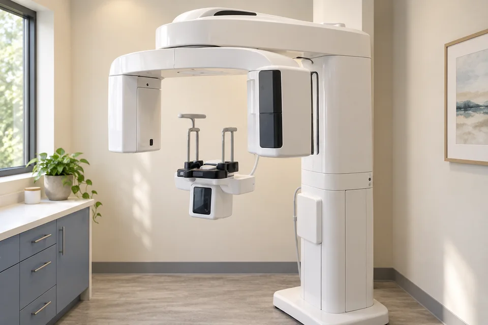
When needed, cone-beam CT (CBCT) gives a 3D view of the jawbone that a surface scan cannot show. Together with digital tooth scans, it helps us consider the bone, implant position, bite and final tooth.
For full-arch restorations, our intraoral scanner combines digital scanning with implant photogrammetry to record the positions of the implants. These records help us design the teeth that attach to them.
Aligners, retainers and your smile
Digital scans help us explain tooth position, plan clear-aligner treatment, follow changes and make retainers. Conventional impressions may still be needed for some cases.
A treatment simulation lets you see the proposed tooth movements. It does not promise exactly how your teeth will move.
For selected cosmetic, restorative and implant cases, facial scanning helps us consider how your teeth fit your smile and face.
We use those images to discuss possible changes with you. Clinical judgment still guides treatment, and a scan cannot promise an exact cosmetic outcome.
X-rays chosen for your care
We take dental images when they help answer a clinical question or plan treatment. Our portable carbon-nanotube digital X-ray system captures intraoral images in a fraction of a second.
Careful positioning and limiting the beam to the area being examined help avoid unnecessary exposure and retakes. The American Dental Association no longer recommends routine lead aprons or thyroid collars for dental X-rays because shielding can obstruct the image.
Published research on our type of handheld system examined operator exposure and shielding under specific test conditions. It does not establish that the system gives patients a lower dose than every other device.
Understand your options
We will explain what an image or scan shows, how it affects your options and what happens next. Ask us to walk through anything that is unclear.